Written and medically reviewed by Dr. Sumit Kr. Roy, BDS, MDS (Prosthodontics & Implantology) — Associate Professor; leads one of Eastern India’s advanced CAD-CAM digital dental centres
Clinical review by Dr. Surupa Dutta, BDS, MDS (Periodontology & Implantology)
Ask most patients what determines whether a dental implant succeeds and they will say the implant itself — the brand, the material, the price. Ask an implantologist and the answer is different.
It is the position.
An implant placed half a millimetre and a few degrees off plan can still integrate perfectly and still leave you with a crown that is harder to clean, angled awkwardly, or supported by less bone than it should be. An implant placed correctly is a tooth root that behaves like a tooth root, for decades.
Computer-assisted implant surgery (CAIS) exists for exactly this reason: to close the gap between the position planned on a screen and the position achieved in the bone. Over the past decade that gap has narrowed from millimetres to fractions of a millimetre, and the evidence behind it is now substantial rather than promotional.
This article explains what guided implant surgery actually involves, what the published data shows about each method, and how to judge whether it matters for your case.
What “Computer-Assisted” Actually Means
Implant surgery can be delivered four ways. The distinction is not cosmetic — it is the difference between following a plan and improvising against one.
Freehand. The surgeon places the implant using clinical judgement, anatomical landmarks and experience. Planning may still involve a CBCT, but nothing physically constrains the drill.
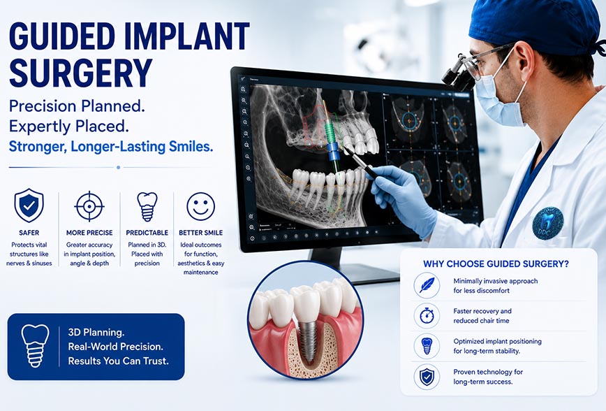
Static CAIS (s-CAIS). The case is planned in 3D, and a surgical guide is designed and 3D-printed. It seats on the teeth, bone or mucosa and holds metal sleeves that control the drill’s entry point, angle and depth. The plan is physically built into the instrument path.
Dynamic CAIS (d-CAIS). Optical tracking follows the handpiece and the patient’s jaw in real time, displaying the drill’s position against the planned position on a screen — a navigation system, conceptually similar to GPS. The surgeon adjusts continuously rather than being constrained mechanically.
Robotic CAIS (r-CAIS). A robotic arm holds the handpiece and physically restricts it to the planned trajectory, depth and angulation. The surgeon operates the system and remains in control; the arm prevents deviation.
All four are performed by a surgeon. None of them make clinical decisions. What separates them is how tightly the execution is bound to the plan.
Why Implant Position Carries So Much Weight
Position determines four things simultaneously:
- Safety. Clearance from the inferior alveolar nerve, mental foramen, maxillary sinus and adjacent roots. A margin of roughly 3 mm between the implant apex and the inferior alveolar nerve is a commonly recommended safety distance — which tells you how quickly a one-millimetre error consumes the available buffer.
- Bone support. Adequate bone must remain around the whole circumference of the fixture. An implant angled towards the buccal plate can perforate thin bone that looked sufficient on a two-dimensional film.
- The final restoration. Prosthetically driven planning means designing the crown first and positioning the implant to serve it. Reverse that order and the crown compensates for the implant — with over-contoured emergence, screw access in the wrong place, or a cement-retained compromise.
- Long-term cleanability. Peri-implantitis, not surgical failure, is the main long-term threat to implant survival. A well-positioned implant is one a patient can actually keep clean.
Guided surgery is not about perfectionism. It is about protecting all four of these at once, in a site where you cannot see what you are drilling into.
How a Guided Case Is Planned
The planning sequence at a digitally equipped clinic looks like this:
- Clinical assessment — bone volume, gum biotype, bite relationship, existing restorations, medical history, smoking and diabetes status
- CBCT imaging — a three-dimensional volume of the jaw showing bone height, width, density and the exact path of nerves and sinuses
- Intraoral digital scanning — an accurate 3D record of the teeth and soft tissue, with no impression trays or setting material
- Data fusion — the CBCT and surface scan are merged into a single working model
- Prosthetic design first — the final crown or bridge is designed in the software before any implant is positioned
- Virtual implant placement — size, depth, angle and emergence chosen against real anatomy, with safety margins verified
- Guide design and manufacture — or export of the plan to a navigation or robotic system
- Guided surgery — the plan is executed as designed
Step 5 is the one patients rarely hear about and the one that most distinguishes a well-planned case. Bone tells you where an implant can go. The crown tells you where it should.
What the Evidence Shows About Accuracy
Accuracy is measured by superimposing a post-operative scan on the original plan and recording three deviations: at the implant platform (coronal), at the tip (apical), and in angulation.
This is one of the better-studied questions in implant dentistry, and independent research groups have converged on remarkably similar answers.
A 2025 systematic review and meta-analysis of robotic computer-assisted implant surgery covering 27 studies, 983 patients and 1,546 implants reported pooled coronal deviation of 0.67 mm, apical deviation of 0.71 mm and angular deviation of 1.68°. Neither the robot system used nor the degree of edentulism significantly affected accuracy, and r-CAIS proved more accurate than freehand, static and dynamic techniques, with only one study reporting an adverse event.
A separate review reached near-identical figures — across 16 clinical studies and 908 implants, average platform deviation was 0.69 mm, apex deviation 0.72 mm and angular deviation 1.62°. A third, published in the prosthodontic literature, found average global coronal, apical and angular deviations of 0.6 mm, 0.7 mm and 1.6 degrees in clinical studies, concluding that robot-assisted placement offered smaller apical and angular deviations than computer-assisted implant surgery.
For comparison, reported deviation values for dynamic navigation have sat around 2.84° angular, 0.75 mm coronal and 1.049 mm apical, with fully guided surgery consistently more accurate than half-guided approaches.
Two things follow from this, and both matter:
Guided beats unguided, consistently. This is the finding with the strongest support and the widest clinical relevance.
The differences between guided methods are real but small. Roughly 0.67 mm versus 0.75 mm at the platform. Decisive beside a nerve in an atrophic mandible; largely academic in a wide posterior site with generous bone. Anyone presenting these differences as transformative for every case is overselling.
The reviews themselves say as much — one notes plainly that both robotic systems and the clinical studies examining them require further development.
Comparing the Four Approaches
| Freehand | Static guided | Dynamic navigation | Robotic | |
| Plan transfer | Surgeon’s judgement | 3D-printed guide | Real-time screen tracking | Physically constrained arm |
| Typical deviation | Highest, most variable | Sub-millimetre in most cases | ~0.75 mm coronal, ~2.8° | ~0.67 mm coronal, ~1.68° |
| Flexibility mid-surgery | Complete | Limited once seated | High | Moderate |
| Irrigation & visibility | Unrestricted | Can be restricted by the guide | Unrestricted | Unrestricted |
| Mouth opening needed | Standard | Extra clearance for sleeve and drill | Standard | Standard |
| Cost to patient | Lowest | Modest addition | Higher | Highest |
| Best suited to | Simple single sites, ample bone | Most routine and multi-unit cases | Cases needing live adjustment | Full-arch and anatomically tight cases |
There is no single correct answer for every patient. A single molar implant in thick bone with 8 mm of clearance above the nerve does not require robotics. A full-arch rehabilitation in a resorbed mandible benefits from every millimetre of control available.
Where AI Fits In — A Supporting Role, Not the Headline
Artificial intelligence enters this workflow mainly at the imaging stage, automating tasks that were previously manual tracing.
A systematic review of U-Net–based deep-learning models for automated CBCT segmentation of the mandibular canal found that these models show strong potential and offer improved efficiency and accuracy, though wider clinical adoption still requires standardised reporting, external validation and explainability to ensure trust and generalisability.
The efficiency gain is the clearest benefit. One study cited in a review of AI in implant planning reported 99.7% accuracy in tooth detection with planning time of about 1.5 seconds compared with 98 seconds for human planning, while another reported detection rates of 72.2% for the mandibular canal and 95.3% for missing teeth. That range — 99.7% down to 72.2% — is the honest summary: performance varies sharply with the structure and the model.
A validation study of AI segmentation of the mandibular incisive canal found a 0.13 mm difference from manual segmentation, unlikely to affect surgical outcomes given the 3 mm safety distance generally recommended, while noting that manual segmentation is considerably more time-consuming.
The review consensus is measured: AI can match or exceed human performance in implant planning, but methodological consistency and ethical compliance still need work before full clinical integration.
In practice: automated segmentation is a fast, consistent first pass that a surgeon then verifies. It saves planning time and reduces the chance of a structure being overlooked at the end of a long day. It does not decide anything, and at our clinic no automated output becomes a surgical plan without clinician confirmation.
How This Works at Digi-Dent Care
We plan implant cases digitally as standard, not as an upgrade:
- Intraoral digital scanning — accurate 3D records without conventional impression trays
- Chairside digital radiography — immediate images at markedly lower radiation than conventional film, with CBCT where the case requires it
- Computer-guided implant planning — prosthetically driven, with the final restoration designed before the implant is positioned
- Guided delivery for single-tooth, multiple-tooth and full-mouth rehabilitation, including immediate implants where indicated
- Implant-supported crowns, bridges and dentures produced through a CAD-CAM workflow
We do not currently operate a surgical robot, and we would rather state that than imply otherwise. On the published evidence, the substantial gain lies in moving from unguided to guided surgery; the step from well-executed guided surgery to robotic surgery is measured in tenths of a millimetre.
What to Ask Before Implant Surgery
- Will my case be planned in 3D from a CBCT, and can I see the plan?
- Is the plan prosthetically driven — designed from the final tooth backwards?
- Will the surgery be guided, and by which method?
- What is the specialist qualification of the person operating?
- What happens if bone volume proves inadequate on the day?
- What does long-term maintenance involve, and what does it cost?
A clinic confident in its workflow will answer all six without hesitation.
The Bottom Line
Computer-assisted implant surgery has moved from a specialist novelty to the reasonable standard of care. Independent meta-analyses show guided placement outperforming freehand consistently, with deviations from plan now well under a millimetre.
What technology cannot do is diagnose, select cases, manage the unexpected, or maintain the implant afterwards. Guided surgery raises the precision ceiling for a good clinician — it does not replace one. The best outcomes still come from an accurate diagnosis, a prosthetically driven plan, a qualified surgeon, and a patient who maintains what has been built.
Digi-Dent Care — Digitally Advanced Multispeciality Dental Clinic, Garia
Computer-guided implant planning · Single, multiple and full-mouth implant rehabilitation · Immediate implants · Implant-supported crowns, bridges and dentures · Intraoral digital scanning · Chairside digital radiography
Address: Saptatari, A/41, New Scheme, Goshtotala, Garia, Kolkata – 700084
Phone: +91 89102 28151
Email: digidentcare24@gmail.com
Book a consultation: www.digidentcare.com/contact-us/
Frequently Asked Questions
1. What is computer-assisted implant surgery?
It is implant placement planned in three dimensions on a computer and then executed through a system that transfers that plan accurately to the mouth — a 3D-printed surgical guide, a real-time navigation system, or a robotic arm. The surgeon performs the surgery throughout; the technology controls how closely the execution matches the plan.
2. Is guided implant surgery better than freehand placement?
For accuracy, yes — this is one of the more consistent findings in the implant literature, with guided techniques outperforming freehand across independent meta-analyses. Experienced surgeons achieve excellent freehand results in favourable sites, but guidance narrows the range of outcomes and matters most where anatomy is tight.
3. How accurate is computer-assisted implant surgery?
Pooled clinical data across nearly 1,000 patients shows deviations from the digital plan of roughly 0.67 mm at the implant platform, 0.71 mm at the tip, and about 1.68° in angulation for robotic systems. Dynamic navigation figures are somewhat higher. These are averages with confidence intervals, not guarantees for any individual case.
4. Which method is best for my case?
It depends on anatomy. A single implant in a wide posterior site with abundant bone is well served by static guided surgery. Full-arch rehabilitation, thin ridges, or sites close to the nerve or sinus benefit most from the tightest available control. Your surgeon should be able to explain why a particular method suits your specific anatomy.
5. Does guided surgery hurt less or heal faster?
Often, indirectly. Precise planning can allow a smaller or flapless approach in suitable cases, which typically means less swelling and a more comfortable recovery. Chair time is frequently shorter. Anaesthesia and biological healing are unchanged — osseointegration still takes months.
6. Does computer-assisted surgery cost more?
Usually a modest amount more than freehand, reflecting CBCT imaging, planning time and guide manufacture. Navigation and robotic systems cost more again. Set against this, better positioning reduces the likelihood of restorative compromise or corrective work later.
7. Do I need a CBCT scan for implant treatment?
For most implant cases, yes. A conventional 2D X-ray shows height but not width, density or the precise path of the nerve. CBCT provides the three-dimensional information that meaningful planning requires. Chairside digital radiography covers the routine imaging, with CBCT used where the case calls for it.
8. Can guided surgery be used if I have bone loss?
Detailed 3D planning is arguably most valuable in these cases, since it defines exactly where usable bone exists and whether grafting or sinus lift procedures are needed first. What planning cannot do is create bone that isn’t there — the grafting and healing sequence still applies.
9. Does a robot perform the surgery in robotic implant placement?
No. The robotic arm holds the handpiece and physically prevents deviation from the planned trajectory. The surgeon controls the procedure, makes every clinical decision, and remains responsible for the outcome. The system constrains movement; it does not exercise judgement.
10. Does Digi-Dent Care use computer-guided implant surgery?
Yes. We plan implant cases using intraoral digital scanning, chairside digital X-rays and CBCT where indicated, with prosthetically driven planning and guided delivery for single, multiple and full-mouth cases. We do not currently use a surgical robot, and we say so plainly rather than implying otherwise.
11. How long does the whole process take?
Planning typically takes one to two appointments including imaging and scanning. Surgery itself is often shorter than freehand placement because the sequence is predetermined. Osseointegration then takes roughly three to six months before the final restoration, depending on bone quality and whether grafting was needed.
12. What matters more — the technology or the surgeon?
The surgeon. Guidance improves how precisely a plan is executed, but the plan itself, case selection, and management of anything unexpected all rest on training and experience. The ideal is a specialist-qualified clinician using good technology, not one compensating for the other.
This article is intended for general information and patient education. It is not a substitute for an in-person consultation, examination, or treatment plan. Individual results vary with anatomy, bone quality and general health. Reviewed by the clinical team at Digi-Dent Care, Garia, Kolkata.
Sources: Systematic review and meta-analysis of robotic computer-assisted implant surgery in clinical dental implant placement (27 studies, 983 patients, 1,546 implants), 2025; systematic review and meta-analysis of R-CAIS accuracy, BMC Oral Health, 2025 (16 studies, 908 implants); accuracy assessment of robot-assisted implant surgery, Journal of Prosthetic Dentistry; accuracy comparison of robot-assisted versus static and dynamic computer-assisted implant surgery; systematic review and meta-analysis of U-Net deep-learning CBCT mandibular canal segmentation; validation study of AI-powered mandibular incisive canal segmentation, Clinical Oral Implants Research, 2025; systematic review on the use of AI in planning dental implant procedures, Dentistry Journal, 2026.