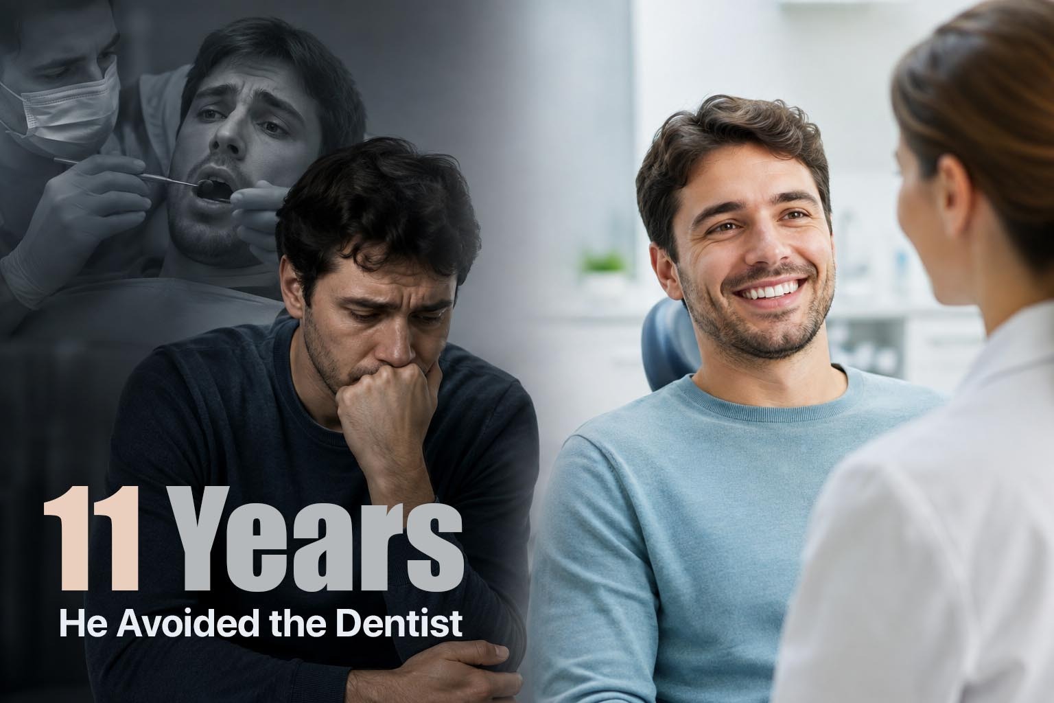
A gentleman in his fifties came to us in Garia with a broken-down upper premolar. Routine enough — the tooth needed a crown.
What was not routine was his reaction when I explained the plan. He went quiet, then asked whether there was any way to do it without “the tray.”
Eleven years earlier, at another clinic, he had gagged so violently during an impression that he vomited in the chair. He described the whole thing to me in detail — the putty pushing back into his throat, the four minutes of waiting, the feeling of not being able to breathe or speak or signal. He had not had restorative dental work since. Two other teeth had broken in the intervening decade. He had simply lived with them.
I told him we would not be using a tray. I picked up the intraoral scanner and showed it to him — a wand about the size of an electric toothbrush handle. I ran it over his lower arch first, so he could watch the model appear on screen before it went anywhere near the part of his mouth he was afraid of.
The scan took about three minutes. He did not gag once.
He looked at the 3D model of his own teeth rotating on the monitor and said something I have heard many times since, in different words: “Nobody told me it had been made this easy.”
That is really the point of this article. Digital intraoral scanning is not a gadget we bought to look modern. It solves problems — for patients and for clinicians — that conventional impressions never fully solved.
What an Intraoral Scanner Actually Is
It is a handheld optical device. A small camera at the tip projects structured light onto your teeth and gums and captures thousands of images per second. Software stitches those images into a single, accurate three-dimensional model of your mouth, which appears on the screen while I am still scanning.
Two clarifications I give patients almost daily:
It is not an X-ray. There is no radiation involved. It is light and a camera — closer in principle to a photograph than to a radiograph.
Nothing is inserted or set. No tray, no putty, no material sitting in your mouth waiting to harden. The wand touches nothing more aggressively than a mirror does, and I can stop and restart at any moment.
For a patient, the experience is: sit, open, follow the wand with your eyes if you like, and it is done in a few minutes.
What Was Wrong With the Old Way
I trained on conventional impressions and took thousands of them. Done well, they produce good results. But there are inherent weaknesses that no amount of skill removes entirely.
The material distorts. Impression material is dimensionally stable within limits, but it shrinks slightly as it sets, it can pull on removal, and it must then be poured into a stone model — every one of those steps introduces a small error. The final crown sits on the end of that chain.
You find out too late. With a conventional impression, I discover a pull, a void or a drag when the material comes out — often after four minutes of the patient’s endurance. If it has failed, we do it again. The patient’s willingness after a second attempt is not always high.
Transport and storage. Impressions travel to a laboratory. They can be distorted in transit, poured incorrectly, or damaged. Stone models chip and get lost.
The patient experience. Gagging, the taste, the pressure, the inability to swallow. For patients with a strong gag reflex, nasal obstruction, anxiety, or limited mouth opening, this is not a minor inconvenience. It is the reason some of them stop coming.
What Changes When the Scan Is Digital
I see the error immediately
This is the single biggest clinical gain, and it is the one patients rarely appreciate.
When I scan a prepared tooth, the model appears on screen at high magnification. If the margin of my preparation is obscured by gingiva or a trace of blood, I see it in that moment — while the patient is still in the chair, still anaesthetised, with the field still isolated. I retract, control the tissue, and rescan that section alone.
With a conventional impression, the same problem means a new tray, new material, and another four minutes.
The scan also magnifies my own work back at me. Preparations look different at 20x on a screen than they do in the mouth. Digital scanning has made me a more meticulous operator, and I say that as someone who thought his preparations were already good.
The model does not distort
The digital file does not shrink, tear, pull, or get damaged in a courier bag. It goes to the laboratory or to our in-house CAD-CAM unit as data. What I captured is what they receive.
Where this shows most clearly is in marginal fit — how precisely the crown seats against the prepared tooth. A margin that is even slightly open is a trap for plaque, and it is where recurrent decay begins. Fewer steps between mouth and restoration means fewer opportunities for that gap to appear.
Treatment planning becomes visual
I can superimpose the scan on a CBCT, design the final crown before positioning an implant, simulate an orthodontic outcome, or run a Digital Smile Design mock-up.
This matters for implants especially. Planning an implant from the bone forwards produces an implant where bone happens to be. Planning it from the final tooth backwards — which requires an accurate surface scan merged with the CBCT — produces an implant that supports the tooth you actually want. That is prosthetically driven planning, and it is not really possible without a digital surface record.
Patients understand what I am telling them
This one surprised me.
When I describe wear on the lingual surfaces of the lower incisors, most patients nod politely and understand nothing. When I rotate a 3D model on a screen and point at it, they understand immediately. Treatment acceptance improves, not because anyone was persuaded, but because they could finally see what I was looking at.
The record is permanent and comparable
Every scan is stored as data. Two years later I can overlay a new scan on the old one and measure change — recession, wear from grinding, tooth movement after orthodontics. This kind of longitudinal comparison was practically impossible with stone models.
Where I Use Scanning at Digi-Dent Care
- Crowns, bridges and veneers — zirconia, metal-free ceramic, implant-supported
- Implant planning — the surface scan merged with CBCT for prosthetically driven, computer-guided placement
- Clear aligners and orthodontic records — where accuracy across the full arch determines whether the aligners fit
- Digital Smile Design — planning and simulating aesthetic outcomes before treatment begins
- Occlusal analysis and wear monitoring — tracking grinding damage over time
- Baseline records — before major rehabilitation, so we know exactly what we started from
What Scanning Still Can’t Do
I would not trust a clinician who told you the technology has no downsides.
Deep subgingival margins remain challenging. A scanner captures what it can see. If a preparation margin sits well below the gum in a bleeding field, no camera can photograph through blood. Tissue management — retraction cord, haemostasis, patience — matters just as much as it always did. Occasionally a conventional impression is still the better tool for a particular margin, and I will use one.
Long fully edentulous spans are harder. Scanning relies on stitching overlapping images together using distinctive landmarks. A long stretch of featureless gum gives the software less to anchor to, and error can accumulate across the arch. This is an active area of development, and scan bodies and stitching algorithms are improving quickly, but it is a genuine current constraint.
The operator still matters more than the device. Scanning has a technique and a learning curve. A poor scan produces a poor restoration exactly as a poor impression does. The scanner did not make the clinician irrelevant — it changed what the clinician needs to be good at.
It is not automatically cheaper. Scanners and CAD-CAM equipment represent a significant investment. What they reduce is remakes, repeat appointments and adjustment visits — which is where the real cost of poor-fitting work has always sat.
What I Would Tell a Patient Deciding Where to Go
Ask whether the clinic uses an intraoral scanner, and ask what they use it for. A clinic that scans only for aligners is different from one that scans for every fixed restoration and implant case.
Ask whether you can see your scan. You should be able to. It is a record of your own mouth, and looking at it will teach you more about your teeth in two minutes than I can explain in ten.
And if you are someone who has avoided dental treatment because of a bad experience with impressions — the gentleman in my chair had waited eleven years — please know that the specific thing you are afraid of is, in most cases, no longer part of the procedure.
He had all three teeth restored over the following months. At his last review he told me he had booked his wife in as well.
Digi-Dent Care — Digitally Advanced Multispeciality Dental Clinic, Garia
Intraoral digital scanning · Digital Smile Design · Computer-guided implant planning · Chairside digital X-rays · CAD-CAM crowns, bridges and implant restorations
Address: Saptatari, A/41, New Scheme, Goshtotala, Garia, Kolkata – 700084
Phone: +91 89102 28151
Email: digidentcare24@gmail.com
Book a consultation: www.digidentcare.com/contact-us/
Frequently Asked Questions
- What is a digital intraoral scan?
It is a small handheld camera that projects light onto your teeth and gums and captures thousands of images, which software assembles into an accurate 3D model of your mouth. It replaces conventional putty impressions for most procedures and the result appears on screen while the scan is still in progress.
- Does intraoral scanning involve radiation?
No. The scanner uses visible or near-infrared light and a camera — there is no ionising radiation. It is often confused with X-rays because both produce images on a screen, but they are entirely different technologies.
- Does it hurt?
No. The wand rests lightly against the teeth and gums, similar to a dental mirror. There is no pressure, no material and nothing to swallow. Most patients describe it as unremarkable, which is precisely the point.
- How long does a scan take?
A single arch typically takes two to four minutes; a full-mouth scan with the bite usually under ten. Complex or full-arch cases take longer, and the scan can be paused at any moment if you need a break.
- I gag badly with impressions. Will I gag with a scan?
Most patients who gag with conventional impressions tolerate scanning well. There is no tray pressing on the palate, no material flowing towards the throat, and no waiting period — and the scan can be stopped and resumed instantly, which removes the trapped feeling that triggers gagging for many people.
- Is a digital scan more accurate than a conventional impression?
For most single-unit and short-span work, digital scanning matches or exceeds conventional impressions, largely by removing steps where distortion occurs — material shrinkage, tray removal, transport and model pouring. For very long fully edentulous spans, conventional techniques still have a role in some situations.
- Can it be used for dental implants?
Yes, and it is particularly valuable there. The surface scan is merged with a CBCT so the final crown can be designed before the implant position is chosen — prosthetically driven planning. It is also used to record implant positions afterwards for the final restoration.
- Can I see my own scan?
Yes, and we encourage it. Seeing a magnified 3D model of your own teeth makes wear, recession, cracks and old restorations far easier to understand than any verbal explanation.
- Will my treatment be faster?
Frequently, yes. Scans transmit to the laboratory instantly rather than by courier, remakes from distorted impressions are largely eliminated, and fewer adjustment appointments are usually needed. Biological healing times — after extractions or implant placement — are unchanged.
- Does scanning cost more than a conventional impression?
The equipment investment is substantial, but for most treatments the fee reflects the procedure rather than the recording method. What scanning reduces is the hidden cost of remakes, repeat visits and restorations that fit imperfectly.
- Are there cases where you would still take a conventional impression?
Occasionally. A preparation margin sitting deep below the gum in a bleeding field, or certain long fully edentulous arches, may still be captured better conventionally. Choosing the right tool for the individual case matters more than using the newest one for everything.
- How is my scan stored, and can it be reused?
It is stored as a digital file in your clinical record. This allows us to compare scans over time to track wear, recession or tooth movement, and to reuse the record for planning further treatment without rescanning unnecessarily.